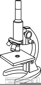
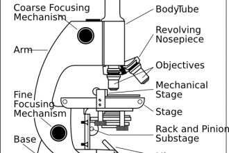
45 labels of a microscope and functions
Confocal Microscopy - an overview | ScienceDirect Topics A confocal microscope was invented in 1951 by Marvin Minsky, a postdoctoral fellow at Harvard University studying neural networks in living brain (Minsky, 1988).In 1957, Minsky patented the concept of confocal imaging, the illumination and detection of a single diffraction-limited spot in a specimen (Fig. 1A).In the transmission configuration, the condenser is replaced with a second … Microscope With Labeled Parts And Functions Microscope With Labeled Parts and Functions. Microscope is a revolutionized scientific instrument which is used in research laboratories to examine the small objects that are not clearly visible and can't be seen by the naked eye. They are derived from Ancient Greek words "mikrós skopeîn" as mikrós means "small" and skopeîn mean "to look" or "see". Microscopes are frequently utilized to research the fungi, microscopic algae,, and other minor life forms.
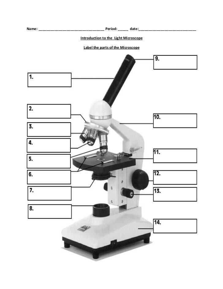
PDF Label parts of the Microscope Label parts of the Microscope: . Created Date: 20150715115425Z

Labels of a microscope and functions
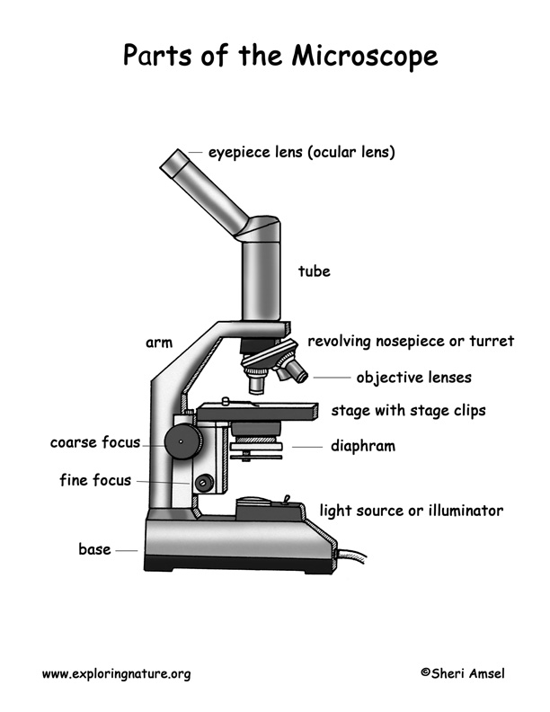
PDF Microscope Parts and their Functions - Landmark Outreach the microscope after you have labeled it on the diagram. 3. After you have completed labeling the diagram, complete the matching exercise. Use the diagram as a guide to help you match the parts of the microscope with their function. a. Write. the letter of the function on the line next to the corresponding microscope part. 4. Microscope Parts and Functions Body tube (Head): The body tube connects the eyepiece to the objective lenses. Arm: The arm connects the body tube to the base of the microscope. Coarse adjustment: Brings the specimen into general focus. Fine adjustment: Fine tunes the focus and increases the detail of the specimen. microscopeinternational.com › compound-microscopeCompound Microscope Parts, Functions, and Labeled Diagram Nov 18, 2020 · Parts of a Compound Microscope Each part of the compound microscope serves its own unique function, with each being important to the function of the scope as a whole. The individual parts of a compound microscope can vary heavily depending on the configuration & applications that the scope is being used for. Common compound microscope parts include: Compound Microscope Definitions for ...
Labels of a microscope and functions. assignmentessays.comAssignment Essays - Best Custom Writing Services Get 24⁄7 customer support help when you place a homework help service order with us. We will guide you on how to place your essay help, proofreading and editing your draft – fixing the grammar, spelling, or formatting of your paper easily and cheaply. Compound Microscope Parts - Labeled Diagram and their Functions - Rs ... The term "compound" refers to the microscope having more than one lens. Basically, compound microscopes generate magnified images through an aligned pair of the objective lens and the ocular lens. In contrast, "simple microscopes" have only one convex lens and function more like glass magnifiers. › 6-label-the-microscopeLabel the microscope — Science Learning Hub Jun 08, 2018 · All microscopes share features in common. In this interactive, you can label the different parts of a microscope. Use this with the Microscope parts activity to help students identify and label the main parts of a microscope and then describe their functions. Drag and drop the text labels onto the microscope diagram. If you want to redo an ... Microscope labeling and functions Flashcards | Quizlet Start studying Microscope labeling and functions. Learn vocabulary, terms, and more with flashcards, games, and other study tools.
Simple Microscope - Parts, Functions, Diagram and Labelling Parts of the optical parts are as follows: Mirror - A simple microscope has a plano-convex mirror and its primary function is to focus the surrounding light on the object being examined. Lens - The biconvex lens is placed above the stage and its function is to magnify the size of the object being examined. A Study of the Microscope and its Functions With a Labeled Diagram These labeled microscope diagrams and the functions of its various parts, attempt to simplify the microscope for you. However, as the saying goes, 'practice makes perfect', here is a blank compound microscope diagram and blank electron microscope diagram to label. Download the diagrams and practice labeling the different parts of these fascinating instruments. Labeling the Parts of the Microscope Labeling the Parts of the Microscope. This activity has been designed for use in homes and schools. Each microscope layout (both blank and the version with answers) are available as PDF downloads. You can view a more in-depth review of each part of the microscope here. Microscope Parts, Function, & Labeled Diagram - slidingmotion Microscope Parts Labeled Diagram The principle of the Microscope gives you an exact reason to use it. It works on the 3 principles. Magnification Resolving Power Numerical Aperture. Parts of Microscope Head Base Arm Eyepiece Lens Eyepiece Tube Objective Lenses Nose Piece Adjustment Knobs Stage Aperture Microscopic Illuminator Condenser Lens
Compound Microscope- Definition, Labeled Diagram, Principle, Parts, Uses The optical microscope often referred to as the light microscope, is a type of microscope that uses visible light and a system of lenses to magnify images of small subjects. There are two basic types of optical microscopes: Simple microscopes. Compound microscopes. The term "compound" in compound microscopes refers to the microscope having ... rsscience.com › stereo-microscopeParts of Stereo Microscope (Dissecting microscope) - Rs' Science Unlike a compound microscope that offers a flat image, stereo microscopes give the viewer a 3-dimensional image that you can see the texture of a larger specimen. [In this image] Examples of Stereo & Dissecting microscopes. Major microscope brands (Zeiss, Olympus, Nikon, Amscope, Omano, Leica …) all produce stereomicroscopes. Microscope Parts, Functions, and Labeling Flashcards | Quizlet Start studying Microscope Parts, Functions, and Labeling. Learn vocabulary, terms, and more with flashcards, games, and other study tools. Microscope Types (with labeled diagrams) and Functions Electron microscope labeled diagram. The different types of electron microscopes are: Transmission Electron Microscope; Scanning Electron Microscope; Reflection Electron Microscope; Scanning transmission electron microscope; Scanning tunneling microscopy; Electron microscope functions: Semiconductors and Data Storage Industry Failure Analysis; Checking for defects
32 Compound Light Microscope Label - Labels 2021
Compound Microscope Parts, Function, & Diagram - Study.com The base of the compound light microscope is the bottom portion of the compound microscope. It functions to support the entire compound microscope. The base can be set on a table or lab bench, and ...
(22).jpg)
An Ultimate Quiz On Microscope Parts And Functions! - ProProfs Quiz
Types of Microscopes: Definition, Working Principle, Diagram ... Where, D is the least distinct vision; F is the focal length of the convex lens; Simple Microscope Diagram. Principle of Simple Microscope. The working principle of a simple microscope is that when a sample is placed within the focus of the microscope, a virtual, erect and magnified image is obtained at the least distance of distinct vision from the eye that is held at the lens.

Motor Neuron Labeled - Top Label Maker
LAS X Industry Microscope software for Industry | Products Activate all relevant functions (e.g. for illumination settings, camera, measurements) with a few clicks ... Add labels for easy analysis. Apply measurements to several images to determine statistical trend and compare data in measurement templates. ... digital reticules adjust to the magnification or zoom of the microscope. LAS X Live Stream ...
Labelling a Microscope - Labelled diagram
Microscope- Definition, Parts, Functions, Types, Diagram, Uses It is a type of fluorescence microscope that is used to produce 2-D or 3-D images of relatively thick specimens. In this type, the excitation light is focused on a specific spot of sample lying on the focal plane. The focus spot is optically manipulated to scan the entire sample and generate a 3-D image.

Biology 2201
Parts of a microscope with functions and labeled diagram Head - This is also known as the body. It carries the optical parts in the upper part of the microscope. Base - It acts as microscopes support. It also carries microscopic illuminators. Arms - This is the part connecting the base and to the head and the eyepiece tube to the base of the microscope.

All Saints Online: Microscope Part Functions
Parts of Stereo Microscope (Dissecting microscope) – labeled … Unlike a compound microscope that offers a flat image, stereo microscopes give the viewer a 3-dimensional image that you can see the texture of a larger specimen. [In this image] Examples of Stereo & Dissecting microscopes. Major microscope brands (Zeiss, Olympus, Nikon, Amscope, Omano, Leica …) all produce stereomicroscopes.
Compound Microscope Clipart | Free Images at Clker.com - vector clip art online, royalty free ...
Parts of a Microscope Labeling Activity - Storyboard That In order to carry out an investigation to show that living things are made of cells you need to be able to use a microscope. Knowing the names of the different parts of the microscope is essential to be able to use one properly. Create a poster that labels the parts of a microscope and includes descriptions of what each part does.
Microscope Unlabelled Diagram - Micropedia
Expansion microscopy - Science Jan 30, 2015 · The resolution of a light microscope is limited. Physicists have long since worked out what these limits are and which parameters determine the spatial resolution. ... resulting in physical magnification. By covalently anchoring specific labels located within the specimen directly to the polymer network, labels spaced closer than the optical ...
35 Drag The Label To The Appropriate Part Of The Microscope. - Labels For Your Ideas
22 Parts Of a Microscope With Their Function And Labeled Diagram Microscopes can broadly be classified into two types: Light microscope and Electron microscope. A light microscope is a type of microscope that commonly uses visible light and a system of lenses to generate magnified images of small objects whereas electron microscope is a microscope that uses a beam of accelerated electrons as a source of illumination.
Microscope With Labels clip art : Biological Science Picture Directory – Pulpbits.net
Microscope, Microscope Parts, Labeled Diagram, and Functions Microscope Parts and their Functions In general, microscopes are made up of supporting parts to ...
Biology label part of microscope
Parts of the Microscope with Labeling (also Free Printouts) Let us take a look at the different parts of microscopes and their respective functions. 1. Eyepiece. it is the topmost part of the microscope. Through the eyepiece, you can visualize the object being studied. Its magnification capacity ranges between 10 and 15 times.
Label a microscope - Teaching resources
Label the microscope — Science Learning Hub Jun 08, 2018 · All microscopes share features in common. In this interactive, you can label the different parts of a microscope. Use this with the Microscope parts activity to help students identify and label the main parts of a microscope and then describe their functions.. Drag and drop the text labels onto the microscope diagram. If you want to redo an answer, click on the …
Label a microscope - Teaching resources
4.1: Cell Structure and Function - Medicine LibreTexts Aug 13, 2020 · A cell is the smallest living thing in the human organism, and all living structures in the human body are made of cells. There are hundreds of different types of cells in the human body, which vary in shape (e.g. round, flat, long and thin, short and thick) and size (e.g. small granule cells of the cerebellum in the brain (4 micrometers), up to the huge oocytes (eggs) …

Vessel Comparison | BioNinja
A Study of the Microscope and its Functions With a Labeled Diagram A Study of the Microscope and its Functions With a Labeled Diagram To better understand the structure and function of a microscope, we need to take a look at the labeled microscope diagrams of the compound and electron microscope. These diagrams clearly explain the functioning of the microscopes along with their respective parts. M mooketsi
Molecular Expressions: Science, Optics & You - Olympus MIC-D: Brightfield Gallery - Spiderwort ...
Parts of a Compound Microscope (And their Functions) List of Microscope Parts and their Functions. 1. Ocular Tubes (Monocular, Binocular & Trinocular) The ocular tubes, are to tubes that lead from the head of the microscope out to your eyes. On the end of the ocular tubes are usually interchangeable eyepieces (commonly 10X and 20X) that increase magnification.

Simple Microscope Labeled Diagram - Micropedia
Two-photon excitation microscopy - Wikipedia Two-photon excitation microscopy (TPEF or 2PEF) is a fluorescence imaging technique that allows imaging of living tissue up to about one millimeter in thickness, with 0.64 μm lateral and 3.35 μm axial spatial resolution. Unlike traditional fluorescence microscopy, in which the excitation wavelength is shorter than the emission wavelength, two-photon excitation requires …

Compound Light Microscope
Classify the structures as homologous or analogous Jun 25, 2018 · The arm of a human and the arm of a chimpanzee have similar functions and bone structures. Frogs and salamanders are both amphibians, and they also both have three-chambered hearts. ... A microscope has a low-power magnification of 200x and a high-power magnification of 1600x. ... 2 Drag each label to the correct location. Not all labels will ...
Post a Comment for "45 labels of a microscope and functions"