45 motor neuron with labels
Motor Neurone Labeled Stock Vector (Royalty Free) 311314823 Find Motor Neurone Labeled stock images in HD and millions of other royalty-free stock photos, illustrations and vectors in the Shutterstock collection. Thousands of new, high-quality pictures added every day. Single-neuron projectome of mouse prefrontal cortex | Nature ... Mar 31, 2022 · a, The pipeline of PFC single-neuron projectome reconstruction including sparse labeling, fMOST imaging, image compression using FNT-slice2cube, neurite tracing using FNT-tracer, quality control ...
Motor Neuron: Function, Types, and Structure | Simply Psychology Motor neurons are responsible for integrating signals from the brain to the muscles, glands, and organs that intend to carry out the required motor function. Motor neurons allow us to move, talk, eat, swallow, and breathe, therefore without these cells, we would not be able to complete many basic life functions.

Motor neuron with labels
Overlapping transcriptional programs promote ... - Neuron Jun 28, 2022 · Several interventions promote survival and regeneration of retinal ganglion cells following injury. scRNA-seq analysis shows that these interventions downregulate a gene expression program associated with cell death and upregulate programs associated with survival and regeneration. Overexpression of some regeneration module genes enhances RGC survival and axon regeneration in vivo. Diagram Quiz on Neuron Structure and Function (Labeling Quiz) Diagram Quiz on DNA replication. 1. Identify the cell type in the above figure. 2. In the figure, labeled '1' receives impulses from adjacent neuron. It is called the. 3. In the figure, labeled '2' is the short filaments from the cell body that carries impulses from dendrites to the cell body which is the. 4. Neuron under Microscope with Labeled Diagram - AnatomyLearner Now, let's discuss the different parts and components of a typical neuron (multipolar motor) with a labelled diagram. But, first, let's try to identify the following features from a neuron with the help of a labelled diagram. Cell body or perikaryon of a neuron Nucleus, cytoplasm, the plasma membrane of a neuron
Motor neuron with labels. Label Neuron Anatomy Printout - EnchantedLearning.com Answers. Neurons. EnchantedLearning.com. Label the Neuron. Brain Glossary. Read the definitions, then label the neuron diagram below. axon - the long extension of a neuron that carries nerve impulses away from the body of the cell. axon terminals - the hair-like ends of the axon. cell body - the cell body of the neuron; it contains the nucleus ... 6.5.2 Draw and label a diagram of the structure of a motor neuron About Press Copyright Contact us Creators Advertise Developers Terms Privacy Policy & Safety How YouTube works Test new features Press Copyright Contact us Creators ... Motor Neuron: Nissl Bodies - Yale University Motor Neuron: Nissl Bodies Nissl stain is used to label rough endoplasmic reticulum in neurons. The dark blue structures are referred to as Nissl bodies but are the equivalent of the rough endoplasmic reticulum. Note how Nissl bodies are confined to the soma and dendrites; they do not extend into the axon. Label a Motor (Multipolar) Neuron Quiz - PurposeGames.com This is an online quiz called Label a Motor (Multipolar) Neuron There is a printable worksheet available for download here so you can take the quiz with pen and paper. This quiz has tags. Click on the tags below to find other quizzes on the same subject. neuron Nervous System Your Skills & Rank Total Points 0 Get started! Today's Rank -- 0
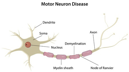
Neuroanatomy, Motor Neuron Article - StatPearls Somatic motor neurons are in the brainstem and further divide into three categories: alpha, beta, and gamma. Alpha motor neurons innervate extrafusal muscle fibers and are the primary means of skeletal muscle contraction. The large alpha motor neuron cell body can be either in the brainstem or spinal cord. In the spinal cord, the cell bodies ... Motor Neuron - The Definitive Guide | Biology Dictionary A motor neuron is a cell of the central nervous system. Motor neurons transmit signals to muscle cells or glands to control their functional output. When these cells are damaged in some way, motor neuron disease can arise. This is characterized by muscle wasting ( atrophy) and loss of motor function. Motor Neuron Overview Neuron Label - The Biology Corner This picture of the neuron is unlabeled, write in the labels to test your knowledge of the anatomy of a neuron. Neuron Label. Publisher: Biologycorner.com; This work is licensed under a Creative Commons Attribution-NonCommercial 3.0 Unported License. This picture of the neuron is unlabeled, write in the labels to test your knowledge of the ... Acetylcholinesterase - Wikipedia Acetylcholinesterase (HGNC symbol ACHE; EC 3.1.1.7; systematic name acetylcholine acetylhydrolase), also known as AChE, AChase or acetylhydrolase, is the primary cholinesterase in the body. It is an enzyme that catalyzes the breakdown of acetylcholine and some other choline esters that function as neurotransmitters: . acetylcholine + H 2 O = choline + acetate
Solved Label the structures of a motor (multipolar) neuron - Chegg Label the structures of a motor (multipolar) neuron by clicking and dragging the labels to the correct location. Dendrites NUcleus Axon collateral Node of Ranvier Synaptic knobs Axon hillock Schwann cell Cell body (soma) N A Labelled Diagram Of Neuron with Detailed Explanations - BYJUS Motor Neurons; The diagram or the structure of the Neuron is useful for both Class 11 and 12 board exams as it has been repetitively asked in the board examinations. It is also one among the few topics having the highest weightage of marks. Learn More: Difference between Sensory and Motor Neuron. Diagram Of Neuron with Labels Single-neuron projectome of mouse prefrontal cortex - Nature 31-03-2022 · Prefrontal cortex (PFC) is the cognitive center that integrates and regulates global brain activity. However, the whole-brain organization of PFC axon projections remains poorly understood. Using ... Motor Neuron - Yale University Motor Neuron Motor Neuron The motor neuron in the ventral horn is easily identifiable by its large size, polygonal shape and extension from the cell body. Compare the size of the nucleolus in the motor neuron with the nuclei in the surrounding support cells. Hide Labels
Anatomy and Physiology 1 Chapter 14 Flashcards | Easy Notecards
Solved DIAGRAM Use the list below to identify and label the - Chegg Question: DIAGRAM Use the list below to identify and label the structures of a motor neuron. Name all 10 parts 4- • • Axon Axon collaterals Axon hillock Axon terminals Cell body or soma Dendrites Myelin sheath Node of Ranvier Schwann cells Trigger zone initial segment) ..HNM von 9 This problem has been solved! See the answer
PPT - Chapter 28 Animal Tissues and Organ Systems (Sections 28.4 - 28.6) PowerPoint Presentation ...
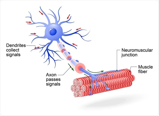
Neuromuscular junction - Wikipedia A neuromuscular junction (or myoneural junction) is a chemical synapse between a motor neuron and a muscle fiber.. It allows the motor neuron to transmit a signal to the muscle fiber, causing muscle contraction.. Muscles require innervation to function—and even just to maintain muscle tone, avoiding atrophy.In the neuromuscular system nerves from the central nervous system and the peripheral ...

Science Review of a Synapse | Free Homework Help
Acetylcholinesterase - Wikipedia Acetylcholinesterase (HGNC symbol ACHE; EC 3.1.1.7; systematic name acetylcholine acetylhydrolase), also known as AChE, AChase or acetylhydrolase, is the primary cholinesterase in the body.

Neuron Cartoon Orange Clip Art at Clker.com - vector clip art online, royalty free & public domain
A Labelled Diagram Of Neuron with Detailed Explanations - BYJUS Motor Neurons The diagram or the structure of the Neuron is useful for both Class 11 and 12 board exams as it has been repetitively asked in the board examinations. It is also one among the few topics having the highest weightage of marks. Learn More: Difference between Sensory and Motor Neuron Diagram Of Neuron with Labels

Pin on Biology of Behavior
Neurofibrillary Tangle - an overview | ScienceDirect Topics Neurofibrillary tangles (NFTs) are most often associated with the neuropathology of CNS neurons affected by Alzheimer's disease and related neurodegenerative disorders, but have also been observed in motor neurons of patients with Alzheimer's disease, 378, 404 with Parkinson dementia complex and amyotrophic lateral sclerosis from Guam, 338, 397 ...

Anterior muscles labeled | Muscle, Labels, Medical
DOC Name: Label the neuron Sensory Neuron - 5. Motor Neuron - 6. Interneuron - 7. Cell Body (Soma) - 8. ... Name: Label the neuron Author: John Girard Last modified by: John Girard Created Date: 5/21/2007 12:41:00 AM Company: Plainville High School Other titles ...

Olig2+ Precursors Produce Abducens Motor Neurons and Oligodendrocytes in the Zebrafish Hindbrain ...
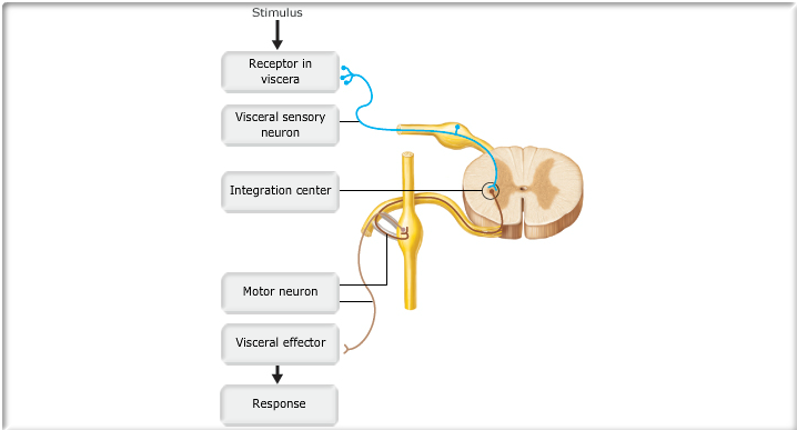
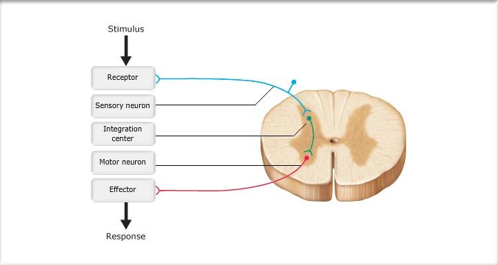
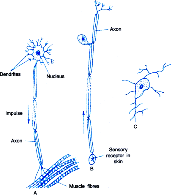
BIO 137 Chapter 11 LS and Quiz Flashcards | Quizlet The sensory neuron conducts information about the stimulus toward the brain or spinal cord. Then, the central nervous system serves as a processing center for the information. The motor neuron then carries an outgoing impulse from the brain or spinal cord. Finally, the effector reacts to stimulation, resulting in the reflex response.
Motor Neuron Disease: Can Dietary Supplements Help?
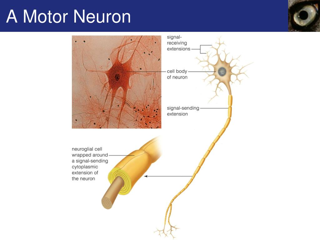
Motor Neuron: Nissl Bodies - medcell.org Motor Neuron: Nissl Bodies Nissl stain is used to label rough endoplasmic reticulum in neurons. The dark blue structures are referred to as Nissl bodies but are the equivalent of the rough endoplasmic reticulum. Note how Nissl bodies are confined to the soma and dendrites; they do not extend into the axon. Hide Labels

http://www.stemcellshealthcare.com/treatable-diseases/motor-neuron-disease-mnd | Motor neuron ...
Motor Neuron - medcell.org Motor Neuron Motor Neuron The motor neuron in the ventral horn is easily identifiable by its large size, polygonal shape and extension from the cell body. Compare the size of the nucleolus in the motor neuron with the nuclei in the surrounding support cells. Hide Labels

Alila Medical Media | Motor neuron unlabeled. | Medical illustration
What color is your…neuron? - NW NOGGIN Aaron Eisen, Outreach Coordinator. What it is: Purkinje Cell. The Purkinje cell is an inhibitory GABAergic neuron which plays an important role in motor control. It is also one of the largest neurons found in the human brain! Purkinje cells are located in the cerebellum, where the majority of all neurons in our brains are found!
Chapter 11 - Neurophysiology Activities Flashcards | Easy Notecards
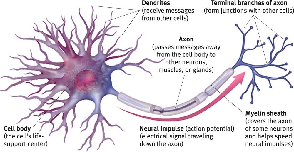
The Brain and Nervous System | Noba Part of the neuron that extends off the soma, splitting several times to connect with other neurons; main output of the neuron. Brain Stem The “trunk” of the brain comprised of the medulla, pons, midbrain, and diencephalon. Broca’s Area An area in the frontal lobe of the left hemisphere. Implicated in language production. Central Nervous ...
Neuron - encyclopedia article - Citizendium
Neurofibrillary Tangle - an overview | ScienceDirect Topics Neurofibrillary tangles (NFTs) are most often associated with the neuropathology of CNS neurons affected by Alzheimer's disease and related neurodegenerative disorders, but have also been observed in motor neurons of patients with Alzheimer's disease, 378, 404 with Parkinson dementia complex and amyotrophic lateral sclerosis from Guam, 338, 397, 473 and with …
What Is Motor Neuron Class 10 | Webmotor.org
Nervous System – Medical Terminology for Healthcare Professions Therefore, this neuron is T-shaped. In the bipolar neuron, the dendrite enters into the left side of the cell body while the axon emerges from the opposite (right) side. In a multipolar neuron, multiple dendrites enter the cell body. The only part of the cell body that does not have dendrites is the part that elongates into the axon.

16 best Motor Neuron Diseases images on Pinterest | Motor neuron, Stem cells and Amyotrophic ...
Label Parts of a Neuron Diagram | Quizlet The chemical signals that neurons use to communicate with other neurons and cells. It is the chemical involved in impulse transmission Neuron transportation Neurons generally transport signals in one direction from the dendrites, through the soma, along the axon and unto the terminal buttons Nodes of Ranvier
Modules 8-14 - PSychology
Neuroanatomy, Motor Neuron - StatPearls - NCBI Bookshelf While the term "motor neuron" evokes the idea that there is only one type of neuron that conducts movement, this is far from the truth. In fact, within the classification of a "motor neuron," there lies both upper and lower motor neurons, which are entirely different in terms of their origins, synapse points, pathways, neurotransmitters, and lesion characteristics. Overall, motor ...

Reading.php Lab
Transcriptomic mapping uncovers Purkinje neuron plasticity ... May 11, 2022 · a, AAV-Plcb4-tdTomato labels Plcb4+ Purkinje neurons transduced by Purkinje neuron-specific GCaMP (AAV-L7-cre + rAAV-CAG-FLEx-jGCaMP7f). Scale bar, 50 μm. Scale bar, 50 μm. Similar results were ...
What is a Neurologic Disorder? - Child Neurology Foundation
Labeled Neuron Diagram | Science Trends Motor neurons are part of the central nervous system (CNS) and communicate signals from the spinal cord to the parts of the body to control their motion. For example, motor neurons send signals to the muscles in your arms causing them to contract. Motor neurons send electrical signals to your intestines so they move and churn food.
Post a Comment for "45 motor neuron with labels"