43 sperm cell diagram with labels
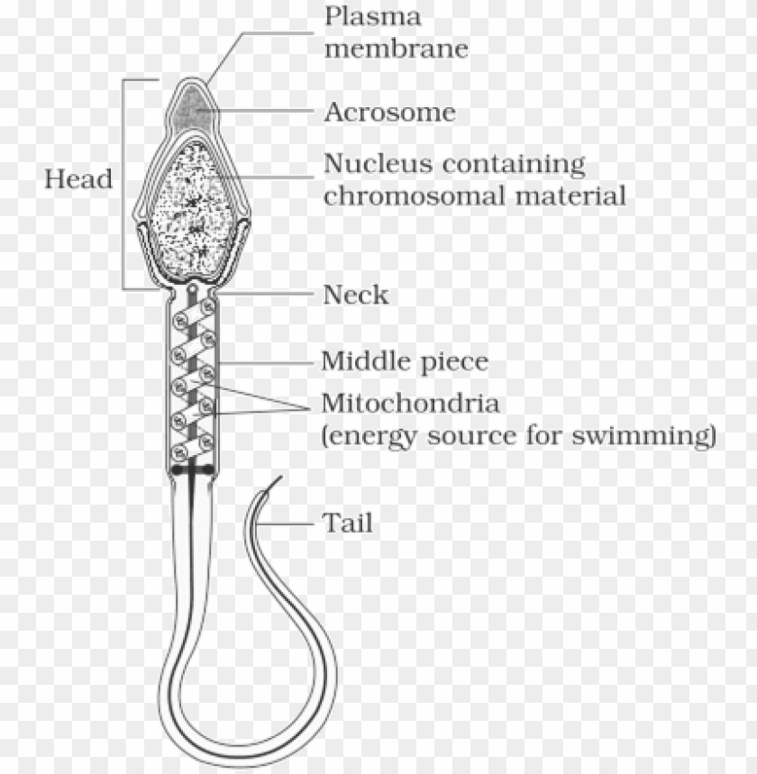
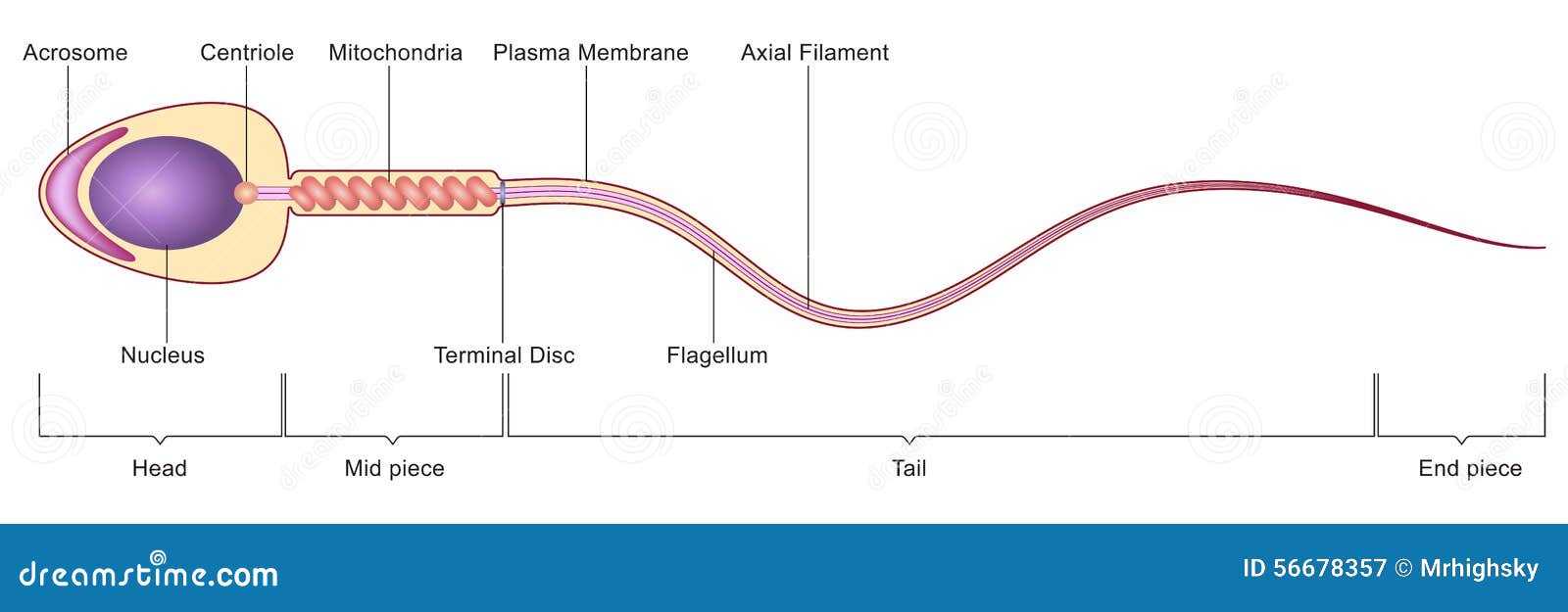
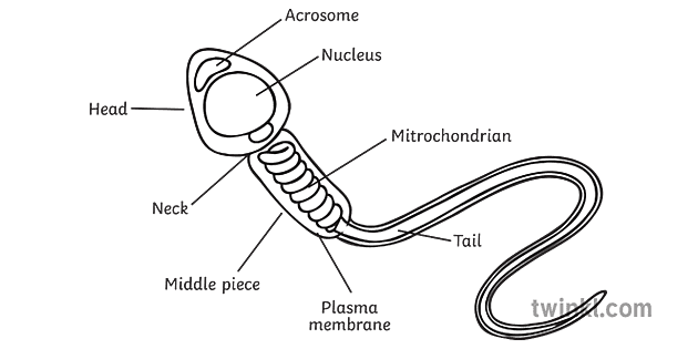
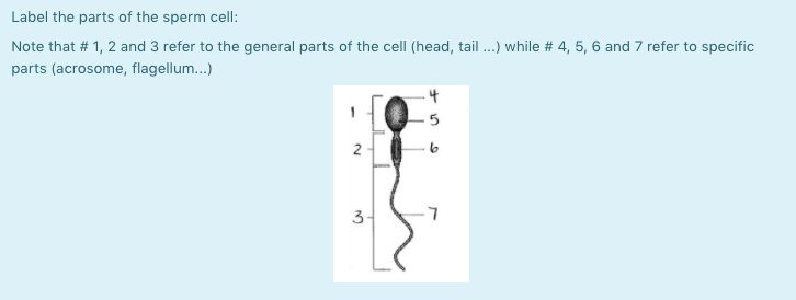
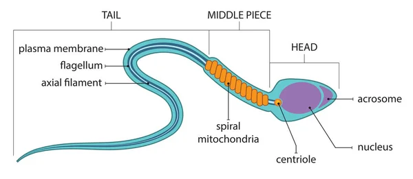
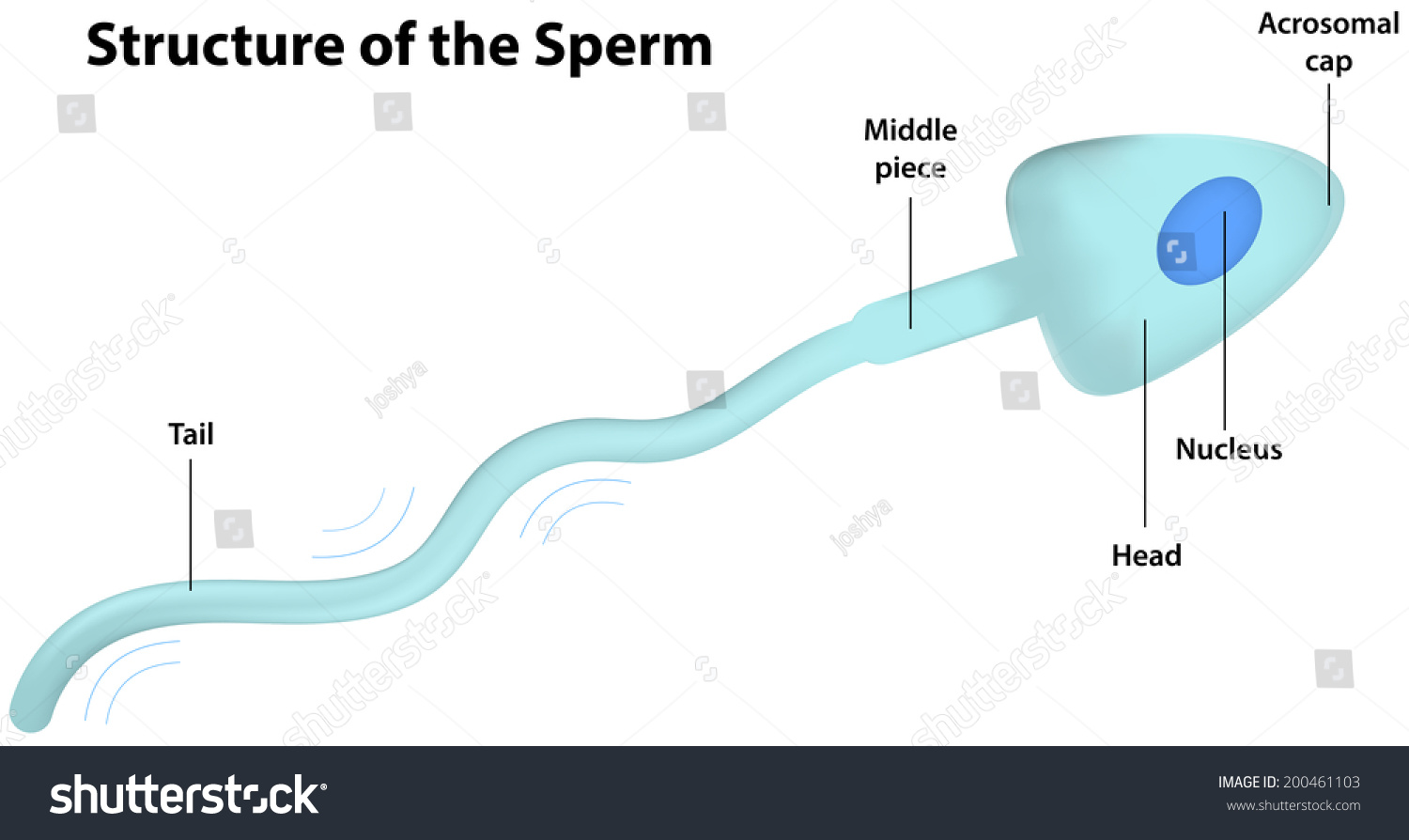
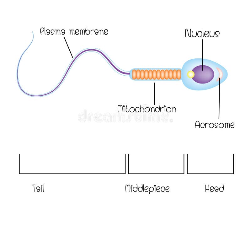
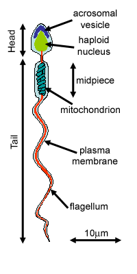
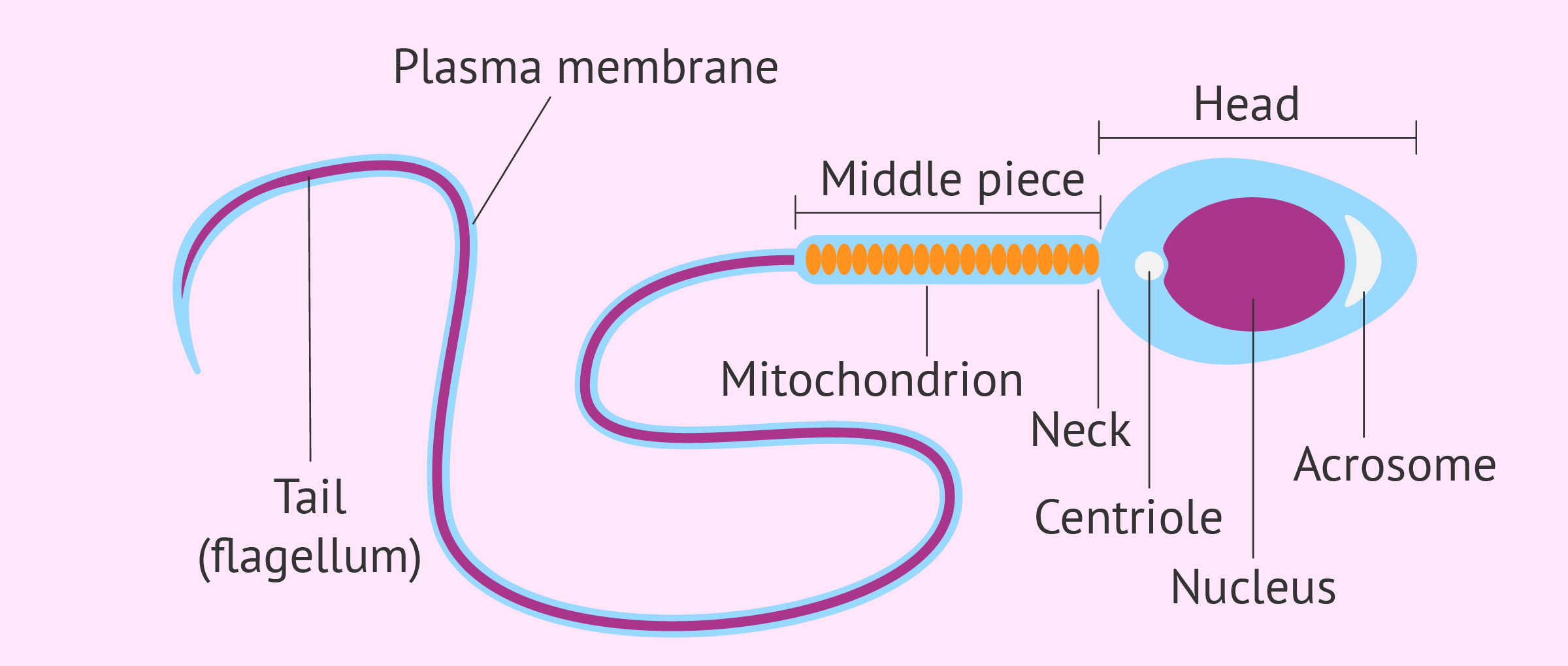
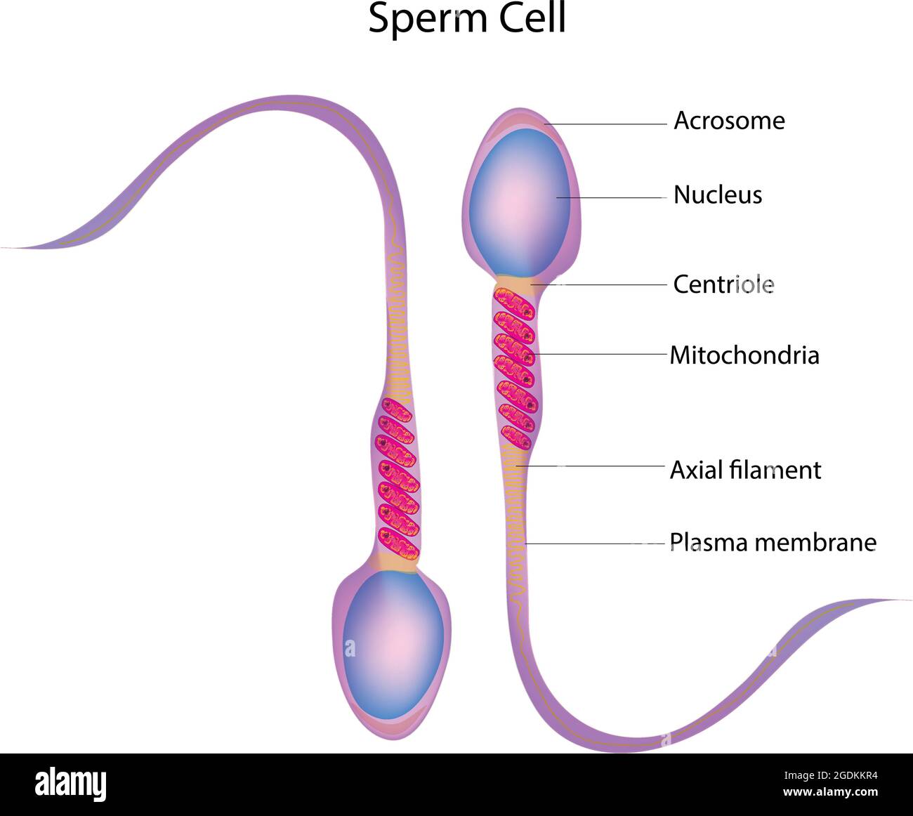
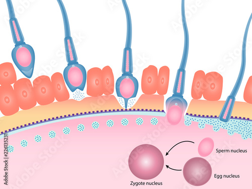
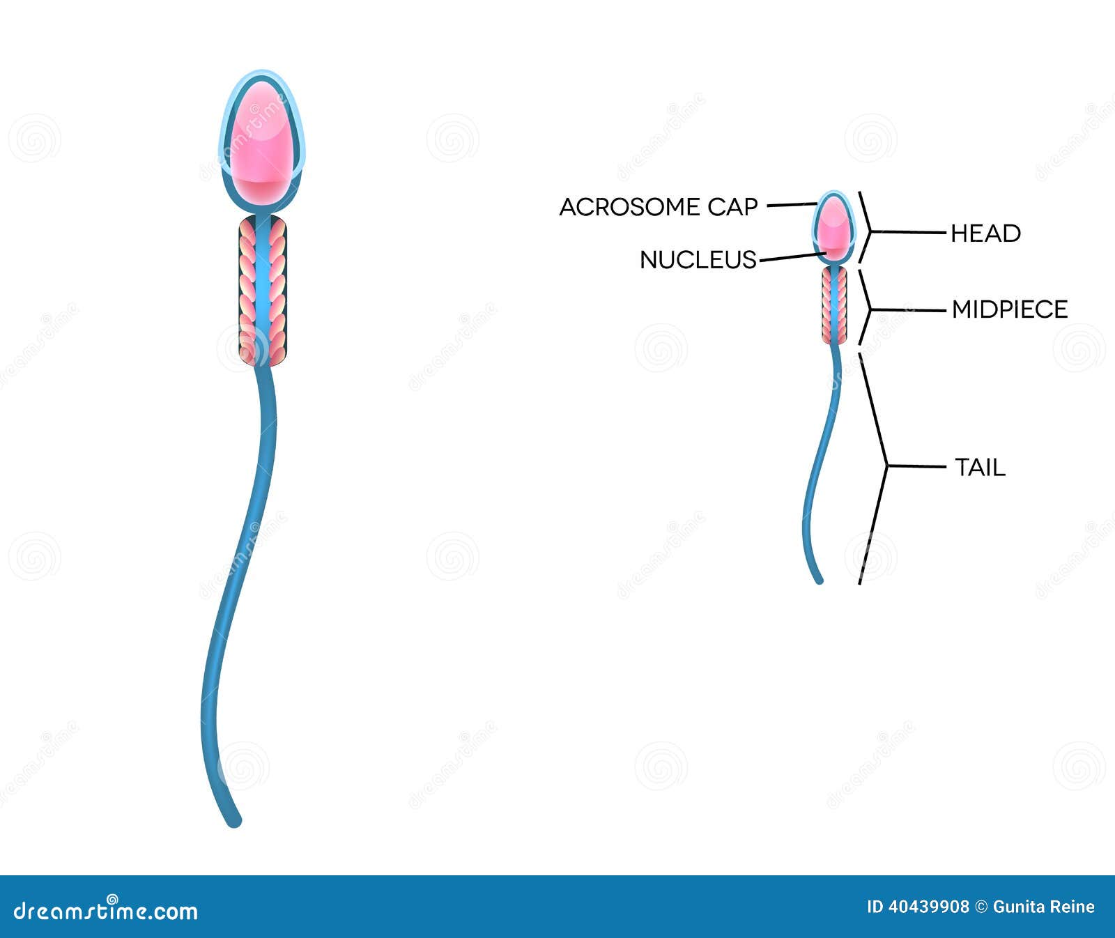
Sperm Under Microscope with Labeled Diagram - AnatomyLearner Sperm Under Microscope with Labeled Diagram 24/06/2022 17/06/2022 by anatomylearner While studying the histological features of the seminiferous tubules and epididymis, you will see sperm cells under the microscope. They are much smaller and lie in groups along the inner margin of the Sertoli cells. Male Reproductive System: Structure, Functions - Embibe The human sperm is microscopic and measures about 40-45 µm in length. Structurally, the sperm is divisible into the head, neck, middle piece and tail. Head The head is a small, almond-shaped structure located at the tip of the sperm that is covered over by a plasma membrane that encloses the acrosome and nucleus.
Animal Cells: Labelled Diagram, Definitions, and Structure - Research Tweet The endoplasmic reticulum (s) are organelles that create a network of membranes that transport substances around the cell. They have phospholipid bilayers. There are two types of ER: the rough ER, and the smooth ER. The rough endoplasmic reticulum is rough because it has ribosomes (which is explained below) attached to it.
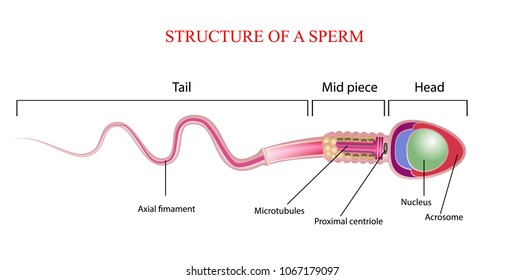
Sperm cell diagram with labels
Structure of Human Sperm: Check Types of Sperm - Embibe - Embibe Exams Explain the Structure of Human Sperm with Labelled Diagram Fig: Structure of a sperm cell Learn Exam Concepts on Embibe What is the Structure of Sperm? Human sperm is a microscopic structure whose shape is like a tadpole. It has flagella which make it motile. Its diameter is \ (2 - 5 {\rm { \mu m}},\) and its length is \ (60 {\rm { \mu m}}.\) Spermatogenesis Diagram & Function | What is the Process of Sperm ... Beneath the Sertoli cells are the spermatogonia, which are germ cells that will go through mitosis and ultimately create sperm. In humans, each day, roughly 25 million spermatogonia divide, and... What is a sperm cell like? Its structure, parts and functions - inviTRA Structure and parts of a sperm cell Neck and middle-piece The neck and the middle piece, as the name suggests, are the parts that can be found between the head and the tail. They measure between 6 - 12 microns, a little longer than the head. The width is hardly visible under the microscope. Inside this part are millions of mitochondria.
Sperm cell diagram with labels. spermatogenesis | Description & Process | Britannica The immature cells (called spermatogonia) are all derived from cells called stem cells in the outer wall of the seminiferous tubules. The stem cells are composed almost entirely of nuclear material. (The nucleus of the cell is the portion containing the chromosomes.) Spermatogenesis- Definition, Stages and Process with figure Spermatocytogenesis is the first stage of spermatogenesis which involves the division of single diploid cells into four haploid spermatocytes. The testis is composed of numerous tightly coiled tubules called seminiferous tubules which are lined with stem cells. The immature cells called spermatogonia are formed from these stem cells. Structure and parts of a sperm cell - inviTRA This labelled diagram shows the structure of a sperm cell in detail, which has the following parts: Head With its spheric shape, it consists of a large nucleus, which at the same time contains an acrosome. The nucleus contains the genetic information and 23 chromosomes. It also secretes a hyaluronidase enzyme that destroys the hyaluronic acid ... sperm | Definition, Function, Life Cycle, & Facts | Britannica sperm, also called spermatozoon, plural spermatozoa, male reproductive cell, produced by most animals. With the exception of nematode worms, decapods (e.g., crayfish), diplopods (e.g., millipedes), and mites, sperm are flagellated; that is, they have a whiplike tail. In higher vertebrates, especially mammals, sperm are produced in the testes. The sperm unites with (fertilizes) an ovum (egg) of ...
Male Reproductive Cell - sperm abstraction abstract bokeh life sex ... Male Reproductive Cell - 16 images - ovarian juvenile granulosa cell tumor human pathology, cervical squamous cell carcinoma human pathology, hyaline cartilage human body help, male reproductive system quiz slide 7, Cell Wall Diagram With Labels - gov.mp Cell Wall Diagram With Labels 1/5 [PDF] Cell Wall Diagram With Labels Printable Animal Cell Diagram - Labeled, Unlabeled, and Blank Unlabeled Animal Cell Diagram Finally, an unlabeled version of the diagram is included at the bottom of the page, in color and black and white. This may be useful as a printable poster for the classroom, or as ... Draw and label the diagram of human sperm cell. Draw and label the diagram of human sperm cell. reproduction; class-10; Share It On Facebook Twitter Email. 1 Answer +1 vote . answered Mar 2 by KalashAtagre (40.7k points) selected Mar 3 by KshitizKumar . Best answer. The diagram of human sperm cell ← Prev Question ... Human Egg Cell Diagram Labeled - embryology of the sea urchin, sperm s ... Human Egg Cell Diagram Labeled - 15 images - human egg cell development, human cells egg stock photo download image now istock, reproductive system worksheet wikieducator, egg cell wikipedia,
Animal Cell- Definition, Structure, Parts, Functions, Labeled Diagram An animal cell is a eukaryotic cell that lacks a cell wall, and it is enclosed by the plasma membrane. The cell organelles are enclosed by the plasma membrane including the cell nucleus. Unlike the animal cell lacking the cell wall, plant cells have a cell wall. Animals are a large group of diverse living organisms that make up three-quarters ... Testes - Anatomy Pictures and Information - Innerbody The testes (singular: testis), commonly known as the testicles, are a pair of ovoid glandular organs that are central to the function of the male reproductive system. The testes are responsible for the production of sperm cells and the male sex hormone testosterone. Cells Diagram | Science Illustration Solutions - Edrawsoft Cells Diagram Symbols Edraw software offers you lots of symbols used in cells diagram like cell structure, paramecium, squamous cell, cell division, bacteria, cell membrane, eggs, sperm, zygote, an animal cell, SARS, tobacco mosaic, adenovirus, coliphage, herpesvirus, AIDS, pollen, plant cell model, onion tissue, etc. Cells Diagram Examples Testes: Anatomy, definition and diagram | Kenhub The testes (testicles) are male reproductive glands found in a saccular extension of the anterior abdominal wall called the scrotum. They are in ovoid shape, sized four to six centimeters in length. Testes develop retroperitoneally on the posterior abdominal wall and descend to scrotum before birth.
Plant Cell: Diagram, Types and Functions - Embibe Exams Q.2. How to make a model of a plant cell diagram step by step procedure? Ans: The plant cell diagram can be checked above and on a similar pattern the diagram can be created. Q.3. Why do plant cells possess large-sized vacuoles? Ans: Vacuole functions in the storage of substances, maintenance of osmolarity and sustaining turgor pressure. Q.4.
Kringe In N Bos Opsommings Graad 11 - 138.68.146.119 Pig Sperm Cell Diagram Labeled.html Solutions For Frq 2014 Physics Ap.html Label The Parts And Blood Vessels Diagram.html Linak Cb9 Service.html Application Format For Transfer Certificate From College.html Reaction Rates And Equilibrium Prentice Hall Key.html E2020 Science Act Diagnostic Test Answers.html Logic Gates Exercises Tdsb.html
What is a sperm cell like? Its structure, parts and functions - inviTRA Structure and parts of a sperm cell Neck and middle-piece The neck and the middle piece, as the name suggests, are the parts that can be found between the head and the tail. They measure between 6 - 12 microns, a little longer than the head. The width is hardly visible under the microscope. Inside this part are millions of mitochondria.
Spermatogenesis Diagram & Function | What is the Process of Sperm ... Beneath the Sertoli cells are the spermatogonia, which are germ cells that will go through mitosis and ultimately create sperm. In humans, each day, roughly 25 million spermatogonia divide, and...
Structure of Human Sperm: Check Types of Sperm - Embibe - Embibe Exams Explain the Structure of Human Sperm with Labelled Diagram Fig: Structure of a sperm cell Learn Exam Concepts on Embibe What is the Structure of Sperm? Human sperm is a microscopic structure whose shape is like a tadpole. It has flagella which make it motile. Its diameter is \ (2 - 5 {\rm { \mu m}},\) and its length is \ (60 {\rm { \mu m}}.\)















Post a Comment for "43 sperm cell diagram with labels"