43 the human heart and its labels
Human Heart Labeling Teaching Resources | Teachers Pay Teachers Human Heart Parts and Blood Flow Labeling Worksheets - Diagram/Graphic Organizer by TechCheck Lessons 4.6 (22) $2.25 Zip This resource contains 2 worksheets for students to (1) label the parts of the human heart and (2) Fill in a flowchart tracing the path of blood flowing though the circulatory system. Answer keys included. How to Draw the Internal Structure of the Heart (with Pictures) - wikiHow To draw the back of the aorta, draw a single line connecting from the right side of the pulmonary artery to the top of the left atrium. To finish drawing the aorta, draw three nubs at the top of the loop. After you draw these, erase the lines connecting from one side of the bottom of the nub to the other. Add tilted circles to the top of all of ...
Human Heart Diagram Without Labels - Pinterest Human Anatomy And Physiology Muscle Anatomy Nursing Notes The heart, blood, and blood vessels are the major components of the cardiovascular system. Like the bustling factory, the body must have a transportation system to carry its various cargos back and forth, and this is where the cardiovascular system steps in. N Nurseslabs

The human heart and its labels
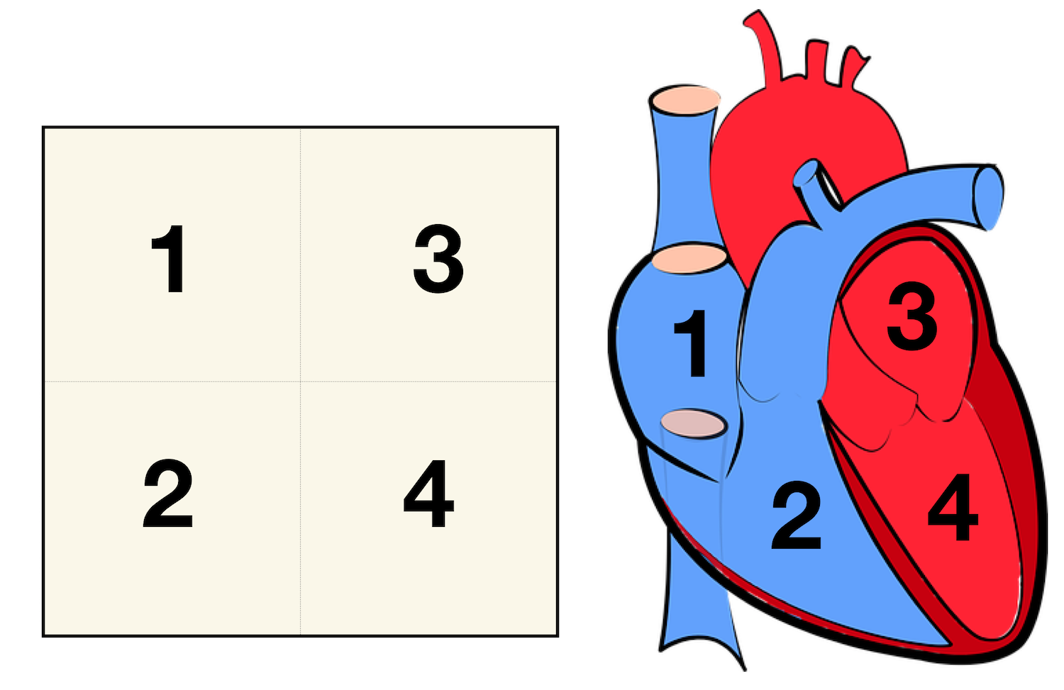
heart | Structure, Function, Diagram, Anatomy, & Facts The heart consists of several layers of a tough muscular wall, the myocardium. A thin layer of tissue, the pericardium, covers the outside, and another layer, the endocardium, lines the inside. The heart cavity is divided down the middle into a right and a left heart, which in turn are subdivided into two chambers. A Diagram of the Heart and Its Functioning Explained in Detail Human heart is covered by a double layered structure which is known as pericardium. The outer layer is associated with the major blood vessels whereas the inner layer is attached to the cardiac muscles. These layers are separated by a pericardial fluid. This covering is like a membrane which holds all the parts of the heart. Chambers Heart Anatomy: Labeled Diagram, Structures, Blood Flow ... - EZmed There are 4 chambers, labeled 1-4 on the diagram below. To help simplify things, we can convert the heart into a square. We will then divide that square into 4 different boxes which will represent the 4 chambers of the heart. The boxes are numbered to correlate with the labeled chambers on the cartoon diagram. View fullsize
The human heart and its labels. Heart Labeled Illustrations & Vectors - Dreamstime Sketch of human heart anatomy ,line and color on a checkered background. Educational diagram with hand written labels of the main. Love Songs Black Golden Silver Records Hearts. Vagus nerve labeled and human organs, medically Illustration. 7 chakras and organs explanation as holistic healing basics outline concept. The Anatomy of the Heart, Its Structures, and Functions - ThoughtCo The heart is made up of four chambers: Atria: Upper two chambers of the heart. Ventricles: Lower two chambers of the heart. Heart Wall The heart wall consists of three layers: Epicardium: The outer layer of the wall of the heart. Myocardium: The muscular middle layer of the wall of the heart. Endocardium: The inner layer of the heart. Human heart: Anatomy, function & facts | Live Science The human heart has four chambers: two upper chambers (the atria) and two lower ones (the ventricles), according to the National Institutes of Health. The right atrium and right ventricle... Heart Diagram with Labels and Detailed Explanation - BYJUS Well-Labelled Diagram of Heart The heart is made up of four chambers: The upper two chambers of the heart are called auricles. The lower two chambers of the heart are called ventricles. The heart wall is made up of three layers: The outer layer of the heart wall is called epicardium. The middle layer of the heart wall is called myocardium.
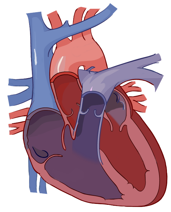
Stock Images, Photos, Vectors, Video, and Music | Shutterstock Stock Images, Photos, Vectors, Video, and Music | Shutterstock Parts Of The Human Heart | Science Trends It is between the lungs, approximately in the middle of the chest, right behind the sternum (breastbone) but slightly to the left. The human heart beats (contracts) each time it received an electrical impulse from the heart muscle, known as the myocardium. The human heart together with the circulatory system make up the cardiovascular system. HOW TO DRAW HUMAN HEART IN VERY EASY STEP - YouTube This is my second video on the Human heart based on general and previous knowledge which we have been reading for years. please go step by step as I am teach... File : Diagram of the human heart (no labels).svg - Wikimedia File:Diagram of the human heart (no labels).svg. From Wikimedia Commons, the free media repository. File. File history. File usage on Commons. Metadata. Size of this PNG preview of this SVG file: 498 × 599 pixels. Other resolutions: 199 × 240 pixels | 399 × 480 pixels | 639 × 768 pixels | 851 × 1,024 pixels | 1,703 × 2,048 pixels | 533 × ...
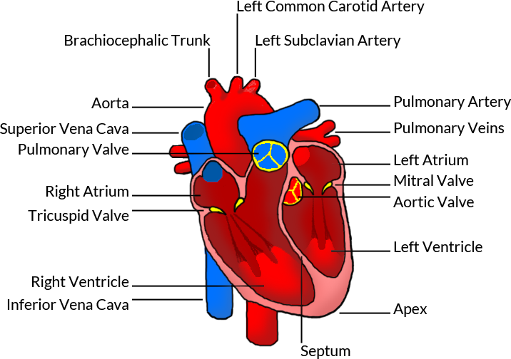
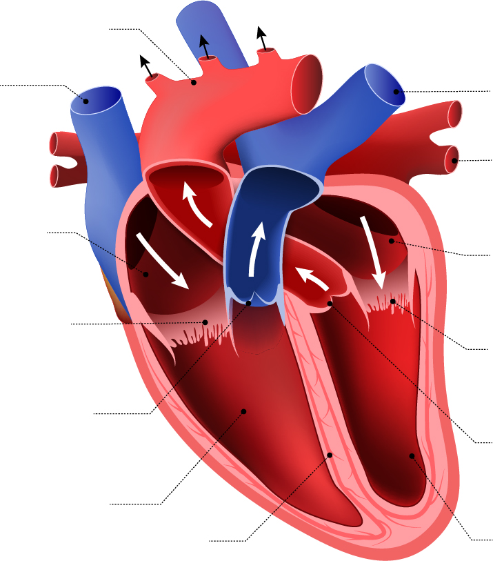
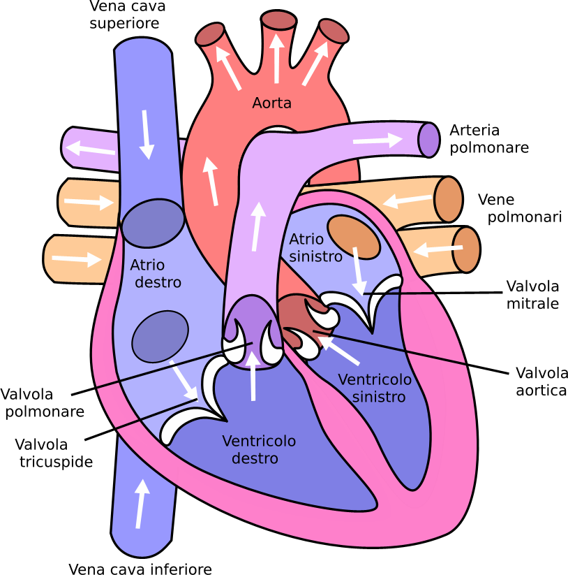
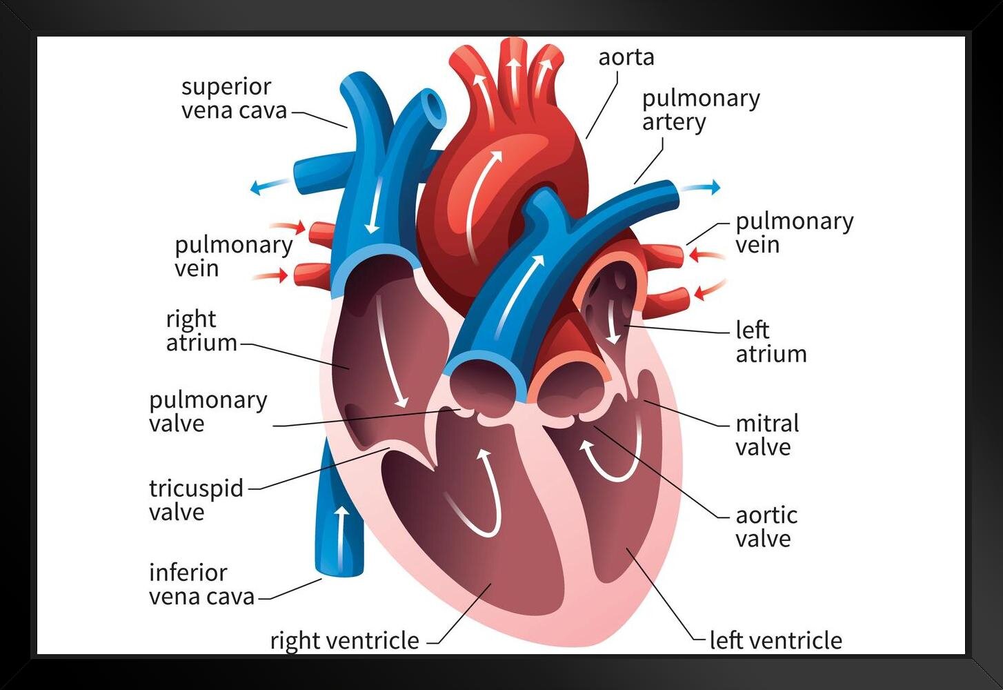
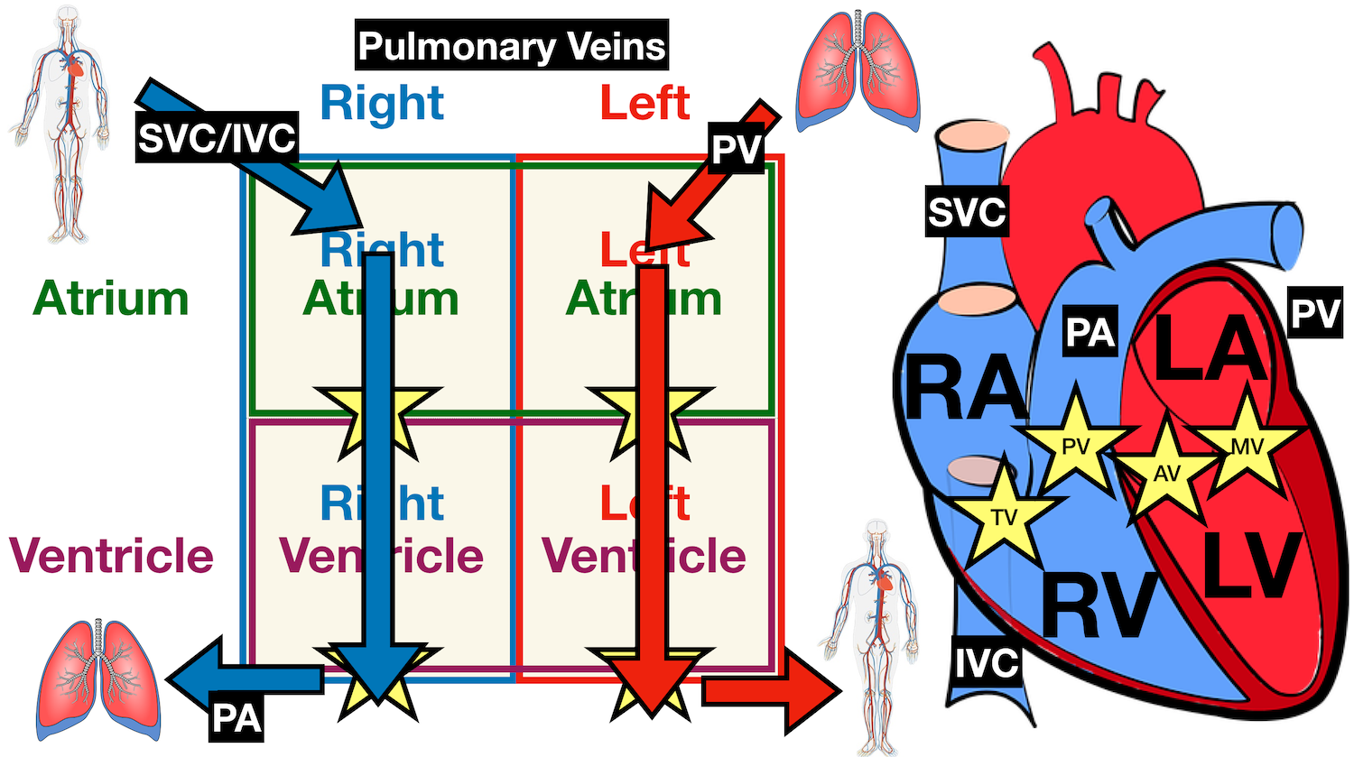
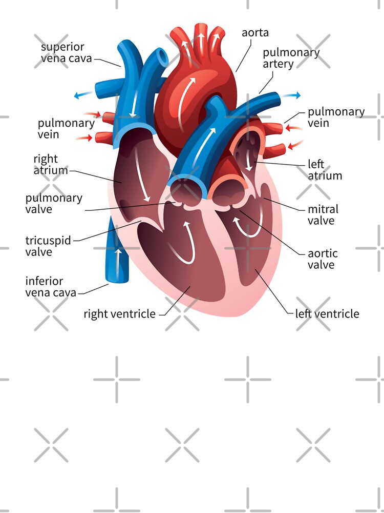
Human Heart - Anatomy, Functions and Facts about Heart - BYJUS The human heart is divided into four chambers, namely two ventricles and two atria. The ventricles are the chambers that pump blood and atrium are the chambers that receive the blood. Among which, the right atrium and ventricle make up the "right portion of the heart", and the left atrium and ventricle make up the "left portion of the heart." 5. Human Heart Diagram Pictures, Images and Stock Photos Browse 4,150 human heart diagram stock photos and images available, or search for heart illustration or pulmonary artery to find more great stock photos and pictures. heart anatomy. Part of the human heart. Human heart cross section, cardiovascular system diagram isolated on white. A Labeled Diagram of the Human Heart You Really Need to See The human heart, comprises four chambers: right atrium, left atrium, right ventricle and left ventricle. The two upper chambers are called the left and the right atria, and the two lower chambers are known as the left and the right ventricles. The two atria and ventricles are separated from each other by a muscle wall called 'septum'. Label the heart — Science Learning Hub In this interactive, you can label parts of the human heart. Drag and drop the text labels onto the boxes next to the diagram. Selecting or hovering over a box will highlight each area in the diagram. pulmonary vein semilunar valve right ventricle right atrium vena cava left atrium pulmonary artery aorta left ventricle Download Exercise Tweet
human heart without label - Clip Art Library
Human Heart Photos and Premium High Res Pictures - Getty Images human heart outline; 21,422 Human Heart Premium High Res Photos. Browse 21,422 human heart stock photos and images available, or search for human heart illustration or human heart icon to find more great stock photos and pictures. heart with arteries and veins - human heart stock pictures, royalty-free photos & images ...
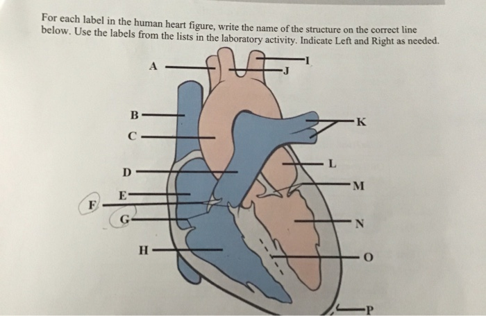
Solved for each label in the human heart figure,write the ...
How to Draw a Human Heart: An Easy Step-By-Step Guide - wikiHow Method 1Sketching the Heart. 1. Draw a tilted and irregular curved shape in the center of your page. Use a pen or pencil to draw the heart's main body. Create a curved shape similar to an acorn or apple's bottom half. Angle the slightly tampered end of the shape to the left about 120 degrees. [1]

Label the heart Diagram | Quizlet
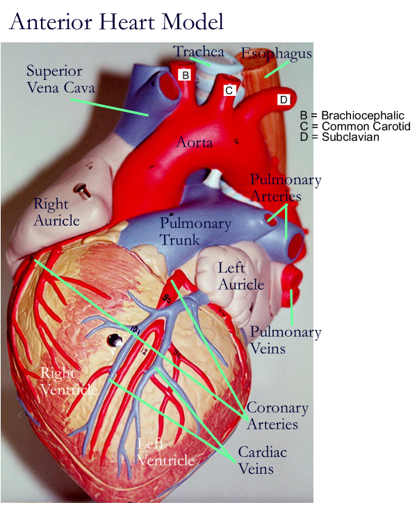
Human Heart Models | Heart Anatomy Models | Vitality Medical The heart model with labels is hand-painted with vivid colors to illustrate the papillary muscles, heart valves, and adjacent structures. Filter by Sort By 4 Items Magnetic Heart Model, Life Size, 5 Part G01 $394.05 View Details Classic Heart Model $81.03 View Details Human Heart Model $450.66 - $566.36 View Details

Draw a diagram of the human heart and label its parts ...
Heart - Wikipedia The human heart is situated in the mediastinum, at the level of thoracic vertebrae T5-T8.A double-membraned sac called the pericardium surrounds the heart and attaches to the mediastinum. The back surface of the heart lies near the vertebral column, and the front surface known as the sternocostal surface sits behind the sternum and rib cartilages. The upper part of the heart is the attachment ...
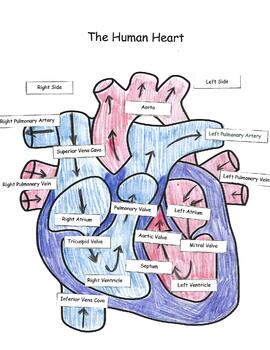
The Human Heart
Human Heart (Anatomy): Diagram, Function, Chambers, Location in Body The heart is a muscular organ about the size of a fist, located just behind and slightly left of the breastbone. The heart pumps blood through the network of arteries and veins called the...

CH. 20 Assessment Flashcards | Quizlet
Human Heart Diagram Labeled | Science Trends The heart has four different chambers: the left and right ventricles and the left and right atriums. The chambers of the heart and the valves that regulate blood flow to them are considered the plumbing of the heart. The left ventricle and left atrium make up the left heart while the right ventricle and right atrium make up the right heart.

Human Heart Diagram Labeled | Science Trends
Heart Diagram with Labels and Detailed Explanation - Collegedunia The human heart is a muscular organ and can be called the most vital organ present in the human body. The heart pumps blood through a network consisting of arteries and veins, a medium also known as the cardiovascular system. The human heart is about the size of a fist, it weighs about 10.5 ounces and it expands and contracts about 100,000 ...
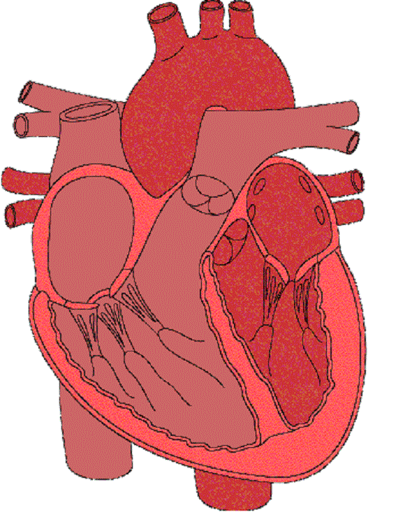
(230).jpg)
Heart Labeling Quiz: How Much You Know About Heart Labeling ...
The 18 parts of the human heart, and their functions The 18 parts of the human heart and how they work 1. Myocardium 2. Endocardium 3. Pericardium 4. Right Auricle 5. Right ventricle 6. Tricuspid valve 7. Pulmonary valve 8. Left Auricle 9. Left ventricle 10. Mitral valve 11. Aortic valve 12. Tendon cords 13. Papillary muscles 14. Sinus node 15. Atrioventricular node 16. Atrioventricular fascicule 17.
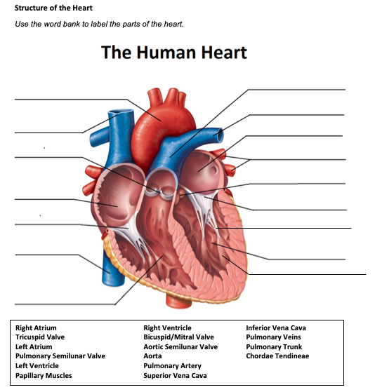
Solved Structure of the Heart Use the word bank to label the ...
Human Heart - Diagram and Anatomy of the Heart - Innerbody The heart contains 4 chambers: the right atrium, left atrium, right ventricle, and left ventricle. The atria are smaller than the ventricles and have thinner, less muscular walls than the ventricles. The atria act as receiving chambers for blood, so they are connected to the veins that carry blood to the heart.
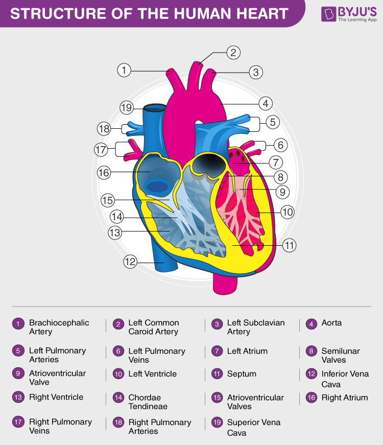
Heart Diagram with Labels and Detailed Explanation
Diagram of Human Heart and Blood Circulation in It A heart diagram labeled will provide plenty of information about the structure of your heart, including the wall of your heart. The wall of the heart has three different layers, such as the Myocardium, the Epicardium, and the Endocardium. Here's more about these three layers. Epicardium

File:Heart diagram-en.svg - Wikimedia Commons
The Human Heart Labeling Worksheet (Teacher-Made) - Twinkl What makes up the anatomy of the human heart? The human heart is a muscle made up of four chambers, these are: Two upper chambers - the left atrium and right atrium Two lower chambers - the left and right ventricles. It's also made up of four valves - these are known as the tricuspid, pulmonary, mitral and aortic valves.

draw and label the structure of a human heart - Brainly.in
Simple Heart Diagram Labeling Activity (Teacher-Made) - Twinkl This simple heart diagram with labels is a fab learning activity to help your pupils aged 10-11 understand the heart and its function in the human body. This simple heart diagram with labels activity will help your pupils begin to understand the heart, what it does and the different parts that comprise it. The resource comes with two different ...
Label the heart — Science Learning Hub
Heart Anatomy: Labeled Diagram, Structures, Blood Flow ... - EZmed There are 4 chambers, labeled 1-4 on the diagram below. To help simplify things, we can convert the heart into a square. We will then divide that square into 4 different boxes which will represent the 4 chambers of the heart. The boxes are numbered to correlate with the labeled chambers on the cartoon diagram. View fullsize
Human Heart Diagram Without Labels - Labelling Worksheet
A Diagram of the Heart and Its Functioning Explained in Detail Human heart is covered by a double layered structure which is known as pericardium. The outer layer is associated with the major blood vessels whereas the inner layer is attached to the cardiac muscles. These layers are separated by a pericardial fluid. This covering is like a membrane which holds all the parts of the heart. Chambers
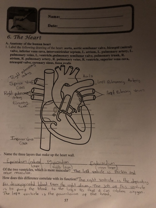
Solved Name: Date: 6. The Heart A. Anatomy of the human ...
heart | Structure, Function, Diagram, Anatomy, & Facts The heart consists of several layers of a tough muscular wall, the myocardium. A thin layer of tissue, the pericardium, covers the outside, and another layer, the endocardium, lines the inside. The heart cavity is divided down the middle into a right and a left heart, which in turn are subdivided into two chambers.

Given alongside is a diagram of the human heart showing its ...
Label the Human Heart | eCampusOntario H5P Studio
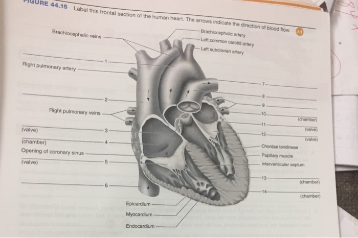
Solved Label this frontal section of the human heart. The ...
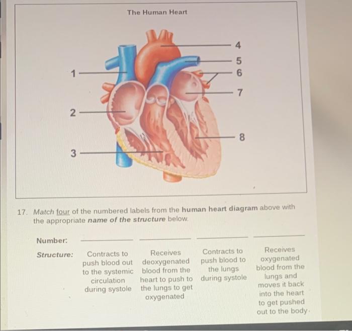
Solved The Human Heart 4 5 6 1 7 N 8 3 17 Match four of the ...
Heart Anatomy: Labeled Diagram, Structures, Blood Flow ...

Simple heart diagram | Simple heart diagram labeled | Human ...

Label the Heart | Heart diagram, Human heart diagram ...

Untitled Document | Human heart diagram, Heart pictures ...
File:Diagram of the human heart (cropped)-it.png - Wikipedia

File:Diagram of the human heart (cropped).svg - Wikimedia Commons
26,637 Human Heart With Labelling Images, Stock Photos ...

Given Alongside is a Diagram of Human Heart Showing Its ...
Human Heart Circulatory System Diagram Chart Medical Educational Science Class Anatomy Corazon Veins Arteries Labels White Wood Framed Art Poster ...
Heart Anatomy: Labeled Diagram, Structures, Blood Flow ...

draw the structure of human heart and label the parts septum ...
The Human Heart: Cut, Paste and Label
poster of human heart anatomy with hand written labels of the ...

Human Body - The Human Heart
Label the heart — Science Learning Hub

draw a diagram of human heart and label its parts? - Brainly.in

Sketch Of Human Heart Anatomy With Hand Written Labels Stock ...

Science worksheets: Label parts of a human heart by Science ...

Human Heart - Anatomy, Functions and Facts about Heart

The Heart - Science Quiz

Human Heart Diagram Class 10 | Get Easy Tricks to Draw Human ...

Pin on MCAT
Heart Models

Human Heart Labeled | Human heart diagram, Heart diagram ...
Post a Comment for "43 the human heart and its labels"