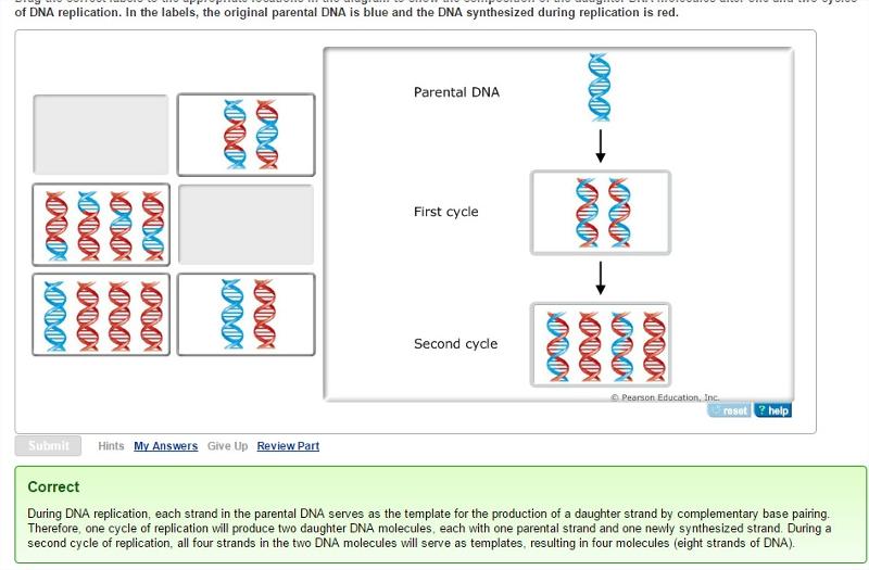
45 diagram of a cell with labels
Plant Cell: Diagram, Types and Functions - Embibe Exams Q.2. How to make a model of a plant cell diagram step by step procedure? Ans: The plant cell diagram can be checked above and on a similar pattern the diagram can be created. Q.3. Why do plant cells possess large-sized vacuoles? Ans: Vacuole functions in the storage of substances, maintenance of osmolarity and sustaining turgor pressure. Q.4. Drawing & Labeling a Diagram of a Electrochemical Cell - Study.com Drawing & Labeling a Diagram of a Electrochemical Cell Instructor: Dave Hays What is an electrochemical cell and what are its component parts? In this lesson, learn what electrochemical cells are,...
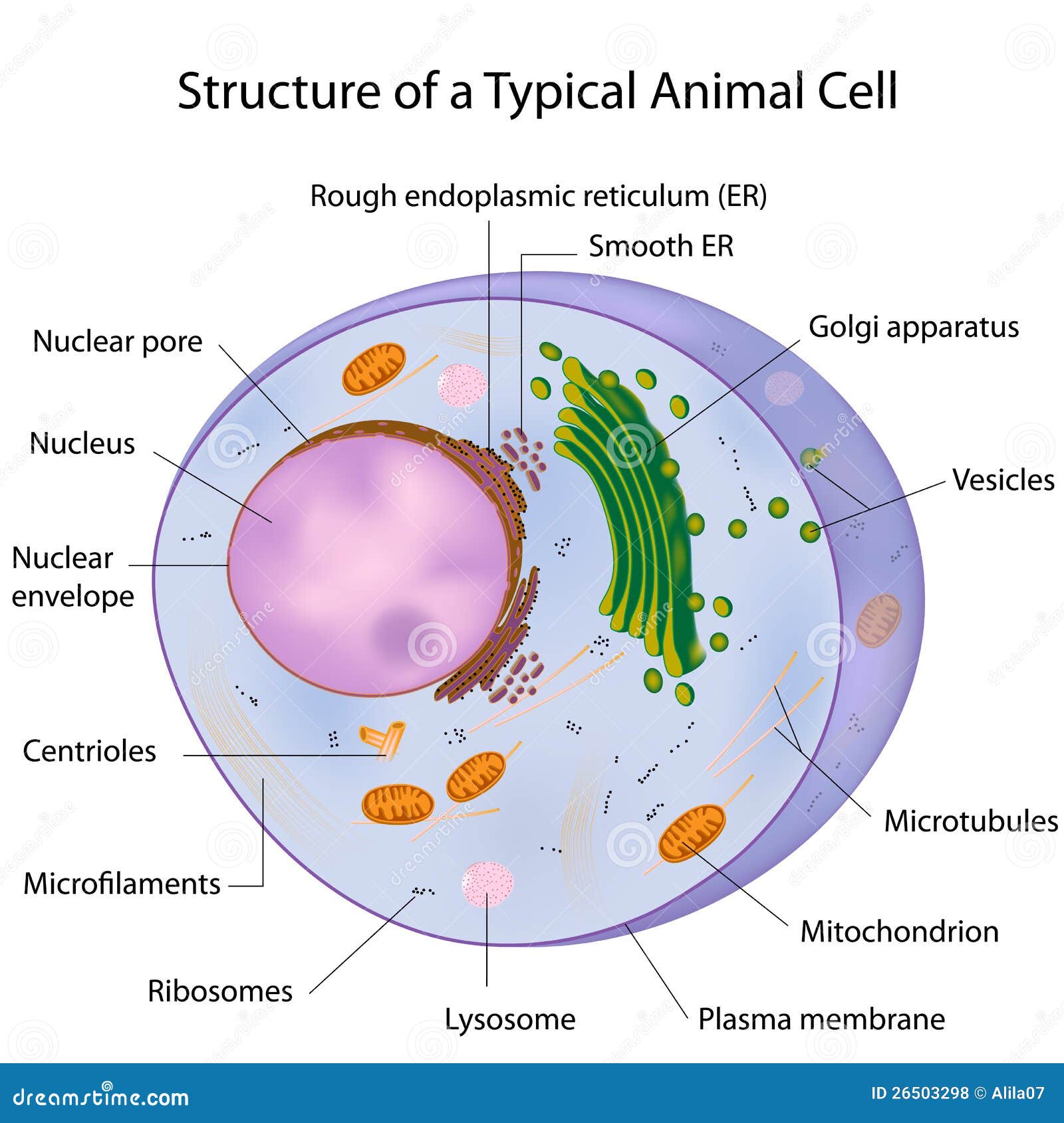
Animal Cell Diagram with Label and Explanation: Cell Structure, Functions Diagram of Animal Cell Below is the diagram of the animal cell which shows the organelles present in it. The cell is covered with cytoplasm which consists of cell organelles in it. The nucleus is covered with a rough Endoplasmic Reticulum and other organelles each designed for a specific purpose.

Diagram of a cell with labels
03 Label the Cell Diagram | Quizlet Start studying 03 Label the Cell. Learn vocabulary, terms, and more with flashcards, games, and other study tools. Animal Cells: Labelled Diagram, Definitions, and Structure Animal Cells Organelles and Functions. A double layer that supports and protects the cell. Allows materials in and out. The control center of the cell. Nucleus contains majority of cell's the DNA. Popularly known as the "Powerhouse". Breaks down food to produce energy in the form of ATP. › cell_cycle_jsInteractive Cell Cycle - CELLS alive INTERPHASE. Gap 0. Gap 1. S Phase. Gap 2. MITOSIS . ^ Cell Cycle Overview Cell Cycle Mitosis > Meiosis > Get the Cell Division PowerPoints
Diagram of a cell with labels. byjus.com › biology › liver-diagramLiver Diagram with Detailed Illustrations and Clear Labels Liver Diagram The liver is one of the most important organs in the human body. Anatomically, the liver is a meaty organ that consists of two large sections called the right and the left lobe. Learn the parts of a cell with diagrams and cell quizzes For this exercise we'll start with an image of a cell diagram ready labeled. Study this and make sure that you're clear about which structure is found where. Cell diagram unlabeled It's time to label the cell yourself! As you fill in the cell structure worksheet, remember the functions of each part of the cell that you learned in the video. Galvanic Cell Diagrams Chemistry Tutorial - AUS-e-TUTE A cell diagram is Chemistry's short-hand for representing a galvanic cell (voltaic cell) . Double vertical lines, || , indicate the connection between the two electrolyte solutions such as a salt bridge. (that is, the oxidation reaction is shown on the left) . When the diagram is read from left to right it shows the direction of the electron ... Structure of Cell: Definition, Types, Diagram, Functions - Embibe Structure of Cell: Cell is the basic functional unit that makes up all living organisms.All organisms, including ourselves, start life as a single cell called the egg. Cells are small microscopic units that perform all essential functions of life and are capable of independent existence.
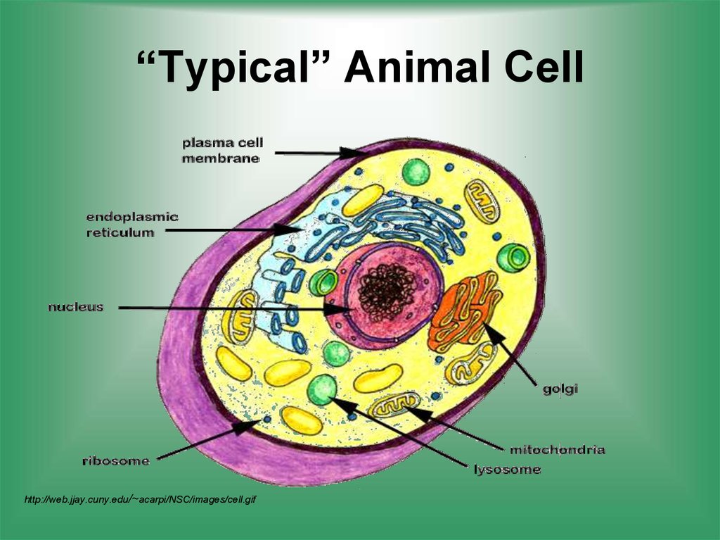
Bacteria in Microbiology - shapes, structure and diagram Bacterial spores. Bacterial endospores layers. Bacteria cells are the smallest living cells that are known; even though viruses are smaller than bacteria, viruses are not living cells. There are different types of bacteria with various sizes, shapes, and structures. The bacteria shapes, structure, and labeled diagrams are discussed below. Animal Cell Diagram | Science Trends The diagram, like the one above, will include labels of the major parts of an animal cell including the cell membrane, nucleus, ribosomes, mitochondria, vesicles, and cytosol. The cells of animals are the basic structural units for the wide variety of life we see in the animal kingdom. Animal cells are eukaryotic in nature, possessing a nucleus ... Animal Cell Diagram - Tim's Printables | Animal cell, Cell diagram ... Plant Cell Labeled Plant Cell Diagram Animal Cell Project Plant And Animal Cells Cell Biology Printable animal cell diagram to help you learn the organelles in an animal cell in preparation for your test or quiz. 5th grade science and biology. S Soul Candy Inter. Design (253) 376-9675 Homeschool Edible Cell Project Plant Cell Project PDF Human Cell Diagram, Parts, Pictures, Structure and Functions Diagram of the human cell illustrating the different parts of the cell. Cell Membrane The cell membraneis the outer coating of the cell and contains the cytoplasm, substances within it and the organelle. It is a double-layered membrane composed of proteins and lipids.
sciencequiz.net › newjcscience › jcbiologyThe Cell - ScienceQuiz.net A is the cell wall and DNA is located inside B. A is the cytoplasm and animal cells may have small vacuoles. A is the cell membrane and B contains chlorophyll. › charts › venn-diagramHow to Create Venn Diagram in Excel – Free Template Download Replace the default values with the custom labels you previously designed. Right-click on any data label and choose “Format Data Labels.” Once the task pane pops up, do the following: Go to the Label Options tab. Click “Value From Cells.” Highlight the corresponding cell from column Label (H2 for Coca-Cola, H3 for Pepsi, and H4 for Dr ... animal and plant cell diagram to label - TeachersPayTeachers 12. $2.00. PDF. Three versions of the plant cell worksheet and three versions of the animal cell worksheet allow students of different grade levels and/or skill levels to label and review the parts of each cell type. Can be used as homework or a quiz. Answer key is provided and can be projected and used during less. How to draw an animal cell - labeled science diagram - YouTube Download a free printable outline of this video and draw along with us: you for watching. Please ...

EPS Vector - A typical cell, labeled, eps10. Stock Clipart Illustration gg62238928 - GoGraph
A Labeled Diagram of the Animal Cell and its Organelles One can observe the golgi apparatus in the labeled animal cell parts diagram. The golgi apparatus is situated near the cell nucleus and besides the stacked sacs, it also contains large number of vesicles. The main function of this golgi complex is to receive the proteins synthesized in the ER and transform it into more complex proteins.

My Classroom
› cells › bactcellInteractive Bacteria Cell Model - CELLS alive In the space are enzymes and other proteins that help digest and move nutrients into the cell. Cell Wall: Composed of peptidoglycan (polysaccharides + protein), the cell wall maintains the overall shape of a bacterial cell. The three primary shapes in bacteria are coccus (spherical), bacillus (rod-shaped) and spirillum (spiral).
Chapter 16- Molecular Basis of Inheritance Flashcards | Easy Notecards
Cells Diagram | Science Illustration Solutions - Edrawsoft Cells Diagram Symbols Edraw software offers you lots of symbols used in cells diagram like cell structure, paramecium, squamous cell, cell division, bacteria, cell membrane, eggs, sperm, zygote, an animal cell, SARS, tobacco mosaic, adenovirus, coliphage, herpesvirus, AIDS, pollen, plant cell model, onion tissue, etc. Cells Diagram Examples
Ch 3 Review Guide
Labeled Plant Cell With Diagrams | Science Trends The parts of a plant cell include the cell wall, the cell membrane, the cytoskeleton or cytoplasm, the nucleus, the Golgi body, the mitochondria, the peroxisome's, the vacuoles, ribosomes, and the endoplasmic reticulum. Parts Of A Plant Cell The Cell Wall Let's start from the outside and work our way inwards.
A Typical Cell, Labeled Stock Photo 112395266 : Shutterstock
Examine the diagram of a cell. Which accurately labels the lysosome? W ... Explanation: Lysosomes are heterogeneous structures present in animal cells which are bound by single membranes. They are of varying shape and size and contains hydrolytic enzymes inside it. Lysosomal membrane has H⁺ ATPase which pumps H⁺ into the membrane through ATP hydrolysis. This pumping of H⁺ makes the internal pH of lysosome acidic.
Cellular Respiration - Kinesthetic Activity | Perkins eLearning
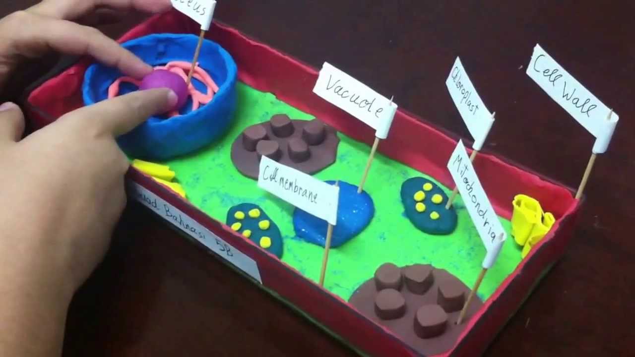
Label Cell Parts | Plant & Animal Cell Activity | StoryboardThat Create a cell diagram with each part of plant and animal cells labeled. Include descriptions of what each organelle does. Click "Start Assignment". Find diagrams of a plant and an animal cell in the Science tab. Using arrows and Textables, label each part of the cell and describe its function.
Describe the structure of nucleus and centrosome with the help of labelled diagram - CBSE Class ...
A Labeled Diagram of the Plant Cell and Functions of its Organelles A Labeled Diagram of the Plant Cell and Functions of its Organelles We are aware that all life stems from a single cell, and that the cell is the most basic unit of all living organisms. The cell being the smallest unit of life, is akin to a tiny room which houses several organs. Here, let's study the plant cell in detail...

Transport in Plants
Cell Membrane With Labels Labeled : Functions and Diagram evmestycor: cell membrane diagram (Virgie Goodwin) There is a printable worksheet available for download here so you can take the quiz with pen and paper. In a plant cell, the cell wall is made up of cellulose, hemicellulose, and proteins while in a fungal cell, it is composed of chitin.

Animal And Plant Cells.
Cell Diagrams - The Biology Corner Open Google Draw and import the diagram. Then use "insert" to create text boxes where students can fill in the labels. Don't forget when assigning this to students on Google classroom to make a copy for each student. You can leave documents in an uneditable form and students can use an addon like "Kami" to annotate the document.
A typical cell, labeled stock vector. Illustration of eukaryote - 26503298
q.uiver.appquiver: a modern commutative diagram editor Edit labels with the input bar at the bottom of the screen. Click and drag the empty space around a object to move it around. Hold Shift (⇧) to select multiple cells to edit them simultaneously.

Draw the general diagram of a plant cell and label it. - Brainly.in
Labeling a Cell Diagram | Quizlet Cell Wall This gives shape and support to the plant cell. It surrounds the cell and protects the other parts of the cell. Chloroplasts This is where the plant cell's chlorophyll is stored. This is what the plant uses to make its own food (photosynthesis). This is also what makes plant cells have a green-like color. Plant cells Are circular in shape

Alila Medical Media | A typical cell, labeled diagram. | Medical illustration
How to draw a nerve cell - labeled science diagrams - YouTube Download a free printable outline of this video and draw along with us: you for watching. Please su...

Alila Medical Media | A typical cell, labeled diagram. | Medical illustration
Cell: Structure and Functions (With Diagram) - Biology Discussion Eukaryotic Cells: 1. Eukaryotes are sophisticated cells with a well defined nucleus and cell organelles. 2. The cells are comparatively larger in size (10-100 μm). 3. Unicellular to multicellular in nature and evolved ~1 billion years ago. 4. The cell membrane is semipermeable and flexible. 5. These cells reproduce both asexually and sexually.
5th grade plant cell project - YouTube
chemostratigraphy.com › how-to-plot-a-ternaryHow to plot a ternary diagram in Excel - Chemostratigraphy.com Feb 13, 2022 · Adding labels to the apices. Next, we need some space for the apices labels: click into the Plot Area (not the Chart Area) then resize by holding the Shift key (this ensures an equal scaling) and use the mouse cursor on one of the corner pick-points. Then recentre the Plot Area in the Chart Area.
pregnancy embryo | Anatomy System - Human Body Anatomy diagram and chart images
A Well-labelled Diagram Of Animal Cell With Explanation Well-Labelled Diagram of Animal Cell The Cell Organelles are membrane-bound, present within the cells. There are various organelles present within the cell and are classified into three categories based on the presence or absence of membrane. Listed below are the Cell Organelles of an animal cell along with their functions.
Cell structure function - презентация онлайн
Plant Cells: Labelled Diagram, Definitions, and Structure Plants have a rigid cell wall that surrounds the plasma membrane. The cell wall is made of cellulose and lignin, which are strong and tough compounds. Plant Cells Labelled Plastids and Chloroplasts Plants make their own food through photosynthesis. Plant cells have plastids, which animal cells don't.
Post a Comment for "45 diagram of a cell with labels"